

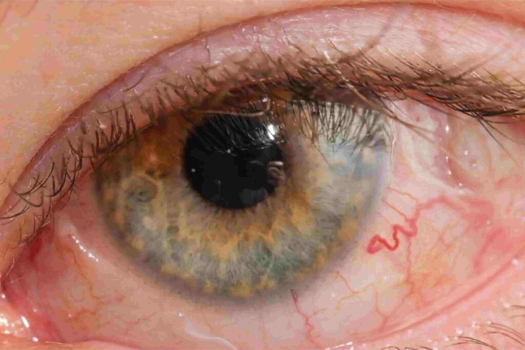
Retinal detachment is a condition in which the retina (the light-sensitive layer at the back of the eye) separates from its underlying support tissue. It is a serious condition that can lead to permanent vision loss if not treated promptly.
Common causes include:
Vitreous haemorrhage is bleeding into the vitreous cavity (the gel inside the eye), which can cause sudden blurring of vision, floaters, or even severe vision loss.
Diagnosis is made through a detailed eye examination, including retinal evaluation. Imaging tests such as OCT or ultrasound may be used when needed.
Retinal detachment usually requires surgery. Treatment options include:
Traditional retinal surgery involves larger instruments and incisions to access the retina, such as in conventional vitrectomy or scleral buckle procedures.
MIVS (Micro-Incision Vitreoretinal Surgery) is a modern technique that uses very small instruments and tiny incisions to perform retinal surgery.
MIVS uses smaller incisions, causes less tissue trauma, and generally allows faster recovery compared to traditional methods.
Retinal surgery involves repairing the retina by removing vitreous gel, sealing retinal breaks, and reattaching the retina using specialized techniques.
Yes, in some cases, cataract surgery can be combined with retinal surgery, depending on the patient’s condition.
If left untreated, retinal detachment can lead to permanent and irreversible vision loss.
Possible complications include: